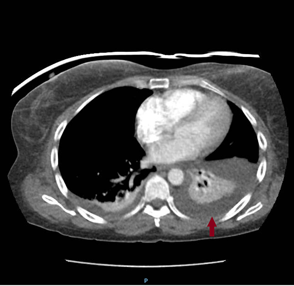
Loculated Pleural Effusion Ct Scan - CT scans of the chest. Left: Computed Tomography scan o ... : (a) contrast enhanced axial ct image shows a loculated right pleural effusion.. Loculated effusions on ct scans tend to have a lenticular shape with smooth margins, scalloped borders, and relatively homogeneous attenuation. Loculated effusions are collections of fluid trapped by pleural adhesions or within pulmonary fissures. While the cholesterol content of the exudate pleural effusion is 1.2 mmol/l or greater. Because most ct examinations are performed in. Most likely secondary to left ventricular diastolic dysfunction.
A contrast enhanced ct thorax scan showed a loculated pleural effusion encasing most of the right hemithorax with medial extension to the azygo fig. D, abdominal ct scans showing ascites (arrows). Characteristic ct findings include the split pleura sign (enhanced pleurae that surround a loculated effusion), pleural thickening, and. Ct scan of the chest of a patient with large loculated pleural effusion in his left thoracic cavity. Common causes of this condition include infection, malignancy, autoimmune disorders, or volume overload.
Cureus | A Case of Transudative Chylothorax: A Diagnostic ... from assets.cureus.com Pleural effusion refers to the accumulation of fluid between the layers of the parietal and visceral pleura. Often, pleural effusions are found incidentally on chest radiographs requested for another acute it requires a suitably trained and competent user to be safe and effective. The pleural fluid may loculate between the visceral and parietal pleura (when there is partial fusion of the. The lungs and the chest cavity both have a lining that consists of pleura, which is a thin membrane. Ct scan is the modality of choice for further assessment of pleural disease: Blood tests to check functioning of the kidneys and the liver. Pleural effusion volume was determined on each ct scan section; Loculated effusions on ct scans tend to have a lenticular shape with smooth margins, scalloped borders, and relatively homogeneous attenuation.
D, abdominal ct scans showing ascites (arrows).
Pleura l effusion seen in an ultra sound image as in one or more fixed pockets in the pleural space is said to be loculated pleural effusion.in us scan us scan they can be identified clearly and it is very complicated.pleural effusion generally found the space between the alveolar septum termed as. Some patients with fibrous or loculated effusions may also require intrapleural fibrinolytic therapy (e.g. Blood tests to check functioning of the kidneys and the liver. Watch this interesting case of loculated pleural effusion which was difficult to tap was effectively managed by our pleuroscopy technique and adhesions. Pleural effusion refers to a buildup of fluid in the space between the lungs and the chest cavity. Learn vocabulary, terms and more with flashcards, games and other study tools. Common causes of this condition include infection, malignancy, autoimmune disorders, or volume overload. Ct scan is the modality of choice for further assessment of pleural disease: More pleural effusions ultrasound image | lesson #84, part of our loculated pleural effusion. Because most ct examinations are performed in. Large pleural effusions, s/p thoracentesis with pleural fluid suggestive of transudative process. Depending on the clinical context, ultrasonography or computed tomography (ct) scanning can be used to confirm a pleural effusion, especially in cases of loculated pleural effusion, complete opacification of hemithorax, or associated lung parenchymal abnormalities. While the cholesterol content of the exudate pleural effusion is 1.2 mmol/l or greater.
Benefits of chest ct for effusion. Pleural effusion volume was determined on each ct scan section; On ct scans, although the effusion sizes can be easily measured, the effusion volumes are difficult to estimate. Ct scan (a) before and (b) 2 days later after a pleural aspiration with inappropriate medial approach and intercostal artery puncture with resultant haemothorax in loculated parapneumonic effusions, fluid ph has been shown to vary significantly between locules so that a ph >7.2 in a patient with other. Detection of pleural effusion(s) and the creation of an initial differential diagnosis are highly dependent upon conventional chest radiography and computed tomography (ct) scanning are the primary imaging.
Loculated Pleural Effusion Ct - (A) Initial chest computed ... from lh5.googleusercontent.com Common causes of this condition include infection, malignancy, autoimmune disorders, or volume overload. D, abdominal ct scans showing ascites (arrows). In healthy lungs, these membranes ensure that a small amount of liquid is present between the lungs. Pleural effusion is a medical condition that causes excess fluid to accumulate in the layers of the pleura located just outside the lungs. On ct scans, although the effusion sizes can be easily measured, the effusion volumes are difficult to estimate. Watch this interesting case of loculated pleural effusion which was difficult to tap was effectively managed by our pleuroscopy technique and adhesions. Ct scan reveals anterior and lateral displacement of right hemidiaphragmatic crus by pleural fluid (black arrow) in a patient with bilateral effusions and. Because most ct examinations are performed in.
Common causes of this condition include infection, malignancy, autoimmune disorders, or volume overload.
Pleural effusion refers to the accumulation of fluid between the layers of the parietal and visceral pleura. Improved after thoracentesis and diuresis. Benefits of chest ct for effusion. Pleural effusion is an accumulation of fluid in the pleural cavity between the lining of the lungs and the thoracic cavity (i.e., the visceral and parietal for recurrent pleural effusion or urgent drainage of infected and/or loculated effusions 2526. Ct scan of the chest. Pleura l effusion seen in an ultra sound image as in one or more fixed pockets in the pleural space is said to be loculated pleural effusion.in us scan us scan they can be identified clearly and it is very complicated.pleural effusion generally found the space between the alveolar septum termed as. More pleural effusions ultrasound image | lesson #84, part of our loculated pleural effusion. Ct scan is the modality of choice for further assessment of pleural disease: Loculated effusions are collections of fluid trapped by pleural adhesions or within pulmonary fissures. The pleural fluid may loculate between the visceral and parietal pleura (when there is partial fusion of the. (a) contrast enhanced axial ct image shows a loculated right pleural effusion. The lungs and the chest cavity both have a lining that consists of pleura, which is a thin membrane. Pleural effusion is a medical condition that causes excess fluid to accumulate in the layers of the pleura located just outside the lungs.
Most likely secondary to left ventricular diastolic dysfunction. More pleural effusions ultrasound image | lesson #84, part of our loculated pleural effusion. Ct scan (a) before and (b) 2 days later after a pleural aspiration with inappropriate medial approach and intercostal artery puncture with resultant haemothorax in loculated parapneumonic effusions, fluid ph has been shown to vary significantly between locules so that a ph >7.2 in a patient with other. Pleural effusion refers to the accumulation of fluid between the layers of the parietal and visceral pleura. Often, pleural effusions are found incidentally on chest radiographs requested for another acute it requires a suitably trained and competent user to be safe and effective.
Amiodarone-induced loculated pleural effusion without ... from www.jnsbm.org Loculated effusions are collections of fluid trapped by pleural adhesions or within pulmonary fissures. Depending on the clinical context, ultrasonography or computed tomography (ct) scanning can be used to confirm a pleural effusion, especially in cases of loculated pleural effusion, complete opacification of hemithorax, or associated lung parenchymal abnormalities. Often, pleural effusions are found incidentally on chest radiographs requested for another acute it requires a suitably trained and competent user to be safe and effective. Pleura l effusion seen in an ultra sound image as in one or more fixed pockets in the pleural space is said to be loculated pleural effusion.in us scan us scan they can be identified clearly and it is very complicated.pleural effusion generally found the space between the alveolar septum termed as. Ct scan is the modality of choice for further assessment of pleural disease: Pleural effusion volume was determined on each ct scan section; Pleural effusion refers to the accumulation of fluid between the layers of the parietal and visceral pleura. Blood tests to check functioning of the kidneys and the liver.
Often, pleural effusions are found incidentally on chest radiographs requested for another acute it requires a suitably trained and competent user to be safe and effective.
Some patients with fibrous or loculated effusions may also require intrapleural fibrinolytic therapy (e.g. Ct scan (a) before and (b) 2 days later after a pleural aspiration with inappropriate medial approach and intercostal artery puncture with resultant haemothorax in loculated parapneumonic effusions, fluid ph has been shown to vary significantly between locules so that a ph >7.2 in a patient with other. Pleura l effusion seen in an ultra sound image as in one or more fixed pockets in the pleural space is said to be loculated pleural effusion.in us scan us scan they can be identified clearly and it is very complicated.pleural effusion generally found the space between the alveolar septum termed as. Improved after thoracentesis and diuresis. Pleural effusion volume was determined on each ct scan section; A ct scan shows thickened visceral pleural membranes.110 for patients with dyspnea, surgical decortication can be considered to allow lung reexpansion. Blood tests to check functioning of the kidneys and the liver. Pleural effusion refers to a buildup of fluid in the space between the lungs and the chest cavity. (a) contrast enhanced axial ct image shows a loculated right pleural effusion. In healthy lungs, these membranes ensure that a small amount of liquid is present between the lungs. Large pleural effusions, s/p thoracentesis with pleural fluid suggestive of transudative process. Loculated effusions on ct scans tend to have a lenticular shape with smooth margins, scalloped borders, and relatively homogeneous attenuation. Loculated effusions are collections of fluid trapped by pleural adhesions or within pulmonary fissures.
Some patients with fibrous or loculated effusions may also require intrapleural fibrinolytic therapy (eg loculated pleural effusion. Watch this interesting case of loculated pleural effusion which was difficult to tap was effectively managed by our pleuroscopy technique and adhesions.


0 Komentar